Infrared imaging discovers source of horse’s ongoing back pain: Overriding dorsal spinous processes (Kissing Spines)
Article by Becky Tenges
FIELD REVIEW: A CASE STUDY Chase is a 15-year old Thoroughbred gelding that is used as a Hunter horse and jumps to 2’ 6”. His owner requested infrared thermal imaging to assist with identifying the possible source of his persistent back pain.
With Thermal Imaging, an infrared detecting camera is used to read radiant heat emitted by an object to create a graphic map of the radiant heat coming from the surface. When used to image horses, the thermal image gives a visual depiction of underlying circulatory levels of the body, providing an objective view of the subjective feeling of pain, and providing the ability to ‘see what the horse feels’.
As opposed to anatomic imaging such as x-rays, which pinpoint the anatomic structures affected, thermal imaging is a physiologic imaging modality, meaning that it detects changes in blood flow and metabolism, and has the ability not only to see tissue conditions but also to recognize areas that are indicative of probable anatomical issues, which can then be further assessed with other, anatomic diagnostic tools.
In the case of Chase, for the past several years, he had been experiencing persistent, but intermittent periods of back pain, which at times were quite severe. Prior to using thermal imaging, his owner and trainer had worked diligently to address Chase’ discomfort and to identify and address possible factors contributing to his back soreness, including: working with a new farrier to address hoof health and balance concerns; contacting the saddle-maker of Chase’ custom-made, but 8 year old saddle, for re-assessment and adjustment of fit (which they repeatedly indicated was fine); working with his vet to provide soundness assessments and joint care and also to provide periodic chiropractic adjustments; creating riding and fitness programs designed to increase Chases’ core strength and the use of his hind end; and, finally, providing Chase with periodic bodywork, to relieve tension and restrictions and to increase range of motion.
And, yet, despite this extraordinary attention to finding and relieving the primary source of Chase’s discomfort, his back pain persisted and, actually, became worse.
Chases’ owner turned to Becky Tenges of Equine BodyWorks for a Saddlefit 4 Life® static diagnostic assessment of his saddle (which, despite the saddle manufacturer’s reaffirmations, actually revealed that his saddle did not fit, extends outside the Saddle Support Area and is well beyond the 18th thoracic rib). The saddle fit assessment was immediately followed by an EquineIR™ full-body thermal imaging assessment.
As with all EquineIR™ imaging, Chases’ scan was done by Becky in accordance with simple, yet strict, quality control guidelines relating to horse preparation, imaging location and environment in order to maximize the quality of the images and minimize the existence and/or influence of artifacts (mud, dirt, sunlight, wind, etc). Chase was imaged at his barn location, eliminating any need for him to travel, leaving him relaxed and stress-free in his own surroundings.
The full-body imaging, which took approximately 30 minutes, included taking 30 strategic images of Chases’ body and hooves. The images were then uploaded to the EquineIR™ Network veterinarian and farrier for analysis. Then, a report was reviewed and images interpreted by a Network veterinarian who, as is the case with all Equine IR™ Network members, are Certified Thermographers (Level I or higher) and also Certified in Equine Thermography.

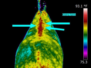
The image seen here is the dorsal view of Chases’ back taken from the rear. Using this and other images from the set of 30 full-body images, the reviewing licensed veterinarian, also a certified thermographer, made the following assessment: “The patterning seen here is consistent with kissing spine (perpendicular hot streaks to thoracic vertebrae at arrows) as well as paraspinal inflammation at the thoracics and lumbars. Radiographs would be useful to confirm OSP [overriding spinous processes] especially given the patient’s history. A saddle-fit scan would be useful, though the report states a static fit confirmed the saddle does not fit appropriately.”
Determining that back pain exists in horses is relatively straightforward. However, accurately determining the source of the pain is confounding, with primary causes which range from hoof to sacroiliac issues, ligament damage to vertebral arthritis, saddle fit to rider-balance issues…to name just a few. The list can feel endless and overwhelming, both emotionally and financially, to an owner.
When Chases’ owner had the benefit of reading the report of the EquineIR™ Network veterinarian, she knew immediately, and with informed confidence, that the right next step was to call her local vet and to incur the cost of radiographs of Chases’ back. The owner made the call. Radiographs were taken and the local veterinarian confirmed the existence of the kissing spine (OSP) that had been revealed and made visible through thermal imaging and the EquineIR™ Network veterinarian’s interpretation.
Of course, this was not a confirmation diagnosis that the owner had hoped to hear, and yet, after years of frustrating efforts to locate the primary source of Chases’ pain, the owner was grateful that thermal imaging had provided the visual clues that permitted her to decide what expense to incur next and his local veterinary team to ‘see where he hurt’ in order to know where to aim their x-ray machine to diagnose his problem.
Thanks to EquineIR™ and thermal imaging, as of the summer of 2013, Chase presently is undergoing treatment for kissing spine. He is on the mend; his pain has subsided considerably; he is under saddle again, lightly and only at the walk; and his owner knows she needs only look to future thermal imaging scans ‘to see what Chases feels’.
Article by Becky Tenges www.equinebodyworksusa.com


